



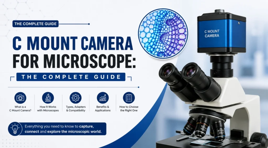
In the world of scientific imaging, few connections are as universally relied upon as the C mount camera for microscope interface. Whether you are outfitting a research laboratory, an industrial quality control station, an academic teaching facility, or a clinical pathology unit, understanding the C mount standard — and choosing the right camera for your microscope — can make the difference between good imaging and truly exceptional imaging. This comprehensive guide explores everything you need to know about C mount microscope cameras, from the basics of the interface to advanced selection criteria and real-world applications.
What Is a C Mount Camera for Microscope?
A C mount camera for microscope is a digital imaging device designed to attach to a microscope via the standardized C mount interface — a 1-inch diameter, 32-thread-per-inch (1″-32 TPI) threaded lens mount that has been an industry standard in machine vision, scientific imaging, and industrial cameras for decades.
The C mount standard was originally developed for 16mm film cameras and later adopted by the broadcast television and closed-circuit television (CCTV) industries. Today, it has become the dominant connector interface for digital cameras used in microscopy, because it provides a rigid, precisely aligned mechanical connection that ensures the camera sensor is correctly positioned at the image plane of the microscope’s optical system.
When a C mount microscope camera is attached to the trinocular port, phototube, or camera port of a microscope, it receives the magnified image formed by the microscope’s objective and eyepiece optical train. The camera’s sensor then converts this optical image into a digital signal that can be displayed on a monitor, captured as a still image, or recorded as a video stream.
Why the C Mount Standard Matters in Microscopy
The precision and rigidity of the C mount connection are not incidental — they are fundamental to image quality. Any mechanical play, misalignment, or tilt between the camera sensor and the microscope’s optical axis introduces aberrations, uneven focus across the field of view, and vignetting (darkening at the image edges). The C mount’s threaded design, when properly tightened and seated, virtually eliminates these problems.
Beyond mechanical alignment, the C mount’s standardization means that a single camera body can be used across a wide variety of microscopes from different manufacturers. This interoperability is enormously valuable in multi-microscope laboratories where the same imaging camera may need to be transferred between instruments.
However, one important nuance to understand is the role of the C mount adapter for microscope — a relay lens system that sits between the microscope’s optical output and the camera’s C mount thread. This adapter serves two purposes: it threads the camera into the microscope port securely, and it adjusts the image magnification (relay factor) so that the circle of microscope image projected onto the camera sensor correctly fills — or nearly fills — the sensor area. Choosing the wrong relay factor adapter can result in either image cut-off (too large an image circle) or wasted sensor area with poor image utilization (too small an image circle).
Key Technical Parameters of a C Mount Microscope Camera
Selecting the right C mount camera for microscope applications requires evaluating several interdependent technical specifications:
1. Sensor Size
Camera sensors for C mount microscopy are available in a range of sizes, from 1/3-inch to 1-inch and beyond. Larger sensors capture more of the microscope’s image field, delivering a wider field of view at the same objective magnification. The sensor size must be matched to the microscope’s phototube design and the C mount adapter’s relay factor.
2. Resolution (Megapixels)
Resolution determines the fineness of detail that can be captured. Entry-level C mount microscopy cameras offer 2–5 megapixels, while professional models provide 12–20 megapixels or more. For applications demanding precise dimensional measurement, documentation for publication, or 4K video output, higher resolution sensors are strongly preferred.
3. Sensor Type: CCD vs CMOS
Charge-Coupled Device (CCD) sensors have long been the gold standard for scientific microscopy due to their low noise, excellent sensitivity, and high dynamic range. However, modern back-illuminated (BSI) CMOS sensors have largely closed this gap and offer additional advantages in speed, frame rate, and cost. For fluorescence microscopy and low-light applications, scientific CMOS (sCMOS) sensors offer exceptional performance.
4. Colour vs. Monochrome
Colour sensors are appropriate for brightfield biological microscopy, histopathology, and any application where colour differentiation is important. Monochrome sensors offer higher sensitivity and resolution for the same pixel count and are preferred for fluorescence imaging, polarized light microscopy, and quantitative measurement tasks.
5. Frame Rate
Live viewing and real-time observation require a minimum of 30 frames per second (fps). High-speed C mount cameras delivering 60 fps or more are available for documenting fast-moving biological processes or high-throughput inspection workflows.
6. Interface and Connectivity
Modern C mount microscopy cameras connect to computers and displays via USB 3.0, USB-C, GigE (Gigabit Ethernet), or Camera Link interfaces. USB 3.0 is the most practical choice for laboratory use due to its widespread compatibility and plug-and-play convenience. WiFi-enabled C mount cameras are increasingly popular for wireless streaming and collaborative observation.
7. Software Compatibility
A professional C mount camera for microscopy should be compatible with industry-standard imaging software such as ImageJ/Fiji, Leica Application Suite, NIS-Elements, and third-party measurement platforms. Open SDK compatibility (often via GenICam or DirectShow) is a major advantage for custom integration.
C Mount vs. CS Mount: What Is the Difference?
A frequent source of confusion in microscopy imaging is the distinction between C mount and CS mount. Both use the same 1-inch, 32 TPI thread, but they differ in the back focal distance — the distance from the mounting flange to the sensor:
- C mount: Back focal distance of 17.526 mm
- CS mount: Back focal distance of 12.526 mm (5 mm shorter than C mount)
Many modern industrial and security cameras use the CS mount. A CS mount camera cannot be used directly on a C mount microscope port without a 5 mm spacer ring — doing so will result in an inability to achieve focus. Conversely, a C mount camera can be used on a CS mount by removing the spacer ring, if one is present. Always verify the mount type of your camera before attempting connection to a microscope port.
Applications of C Mount Cameras for Microscopes
The versatility of the C mount interface means that C mount microscope cameras serve an extraordinarily wide range of professional applications:
Biological Research
Research microscopes used in cell biology, genetics, microbiology, and neuroscience are routinely fitted with high-resolution C mount cameras for fluorescence imaging, time-lapse documentation, and confocal co-localization studies.
Histopathology and Clinical Diagnostics
Pathology laboratories use C mount cameras mounted on upright clinical microscopes to capture and digitize tissue section images for telepathology, digital slide archiving, and second-opinion consultations.
Metallurgical Analysis
Inverted and upright metallurgical microscopes in quality control labs and failure analysis centers are equipped with C mount cameras to document grain structures, corrosion layers, weld cross-sections, and surface defects in metals and alloys.
PCB and Electronics Inspection
Zoom stereo microscopes and inspection microscopes used in electronics manufacturing carry C mount cameras to facilitate solder joint inspection, component placement verification, and defect documentation on printed circuit boards.
Gemology and Jewellery
Gemologists and jewellers use stereo microscopes with C mount cameras to display and photograph inclusions, facet quality, and surface conditions for grading and client communication.
Education and Training
Universities, colleges, and training institutes fit teaching microscopes with C mount cameras to project live microscope images to classroom displays, enabling simultaneous observation by all students and dramatically improving the educational experience.
Tool Maker’s Microscopy
In precision engineering and metrology, tool maker’s microscopes equipped with C mount cameras provide digital documentation of thread profiles, surface finishes, and dimensional measurements.
How to Choose the Right C Mount Adapter for Your Microscope
Selecting the correct C mount adapter for microscope is as important as choosing the camera itself. The adapter must:
- Match the thread or bayonet specification of your microscope’s camera port
- Provide the correct relay factor (0.35×, 0.5×, 0.63×, 1.0×, etc.) to optimally fill the camera sensor
- Be manufactured to tight optical tolerances to avoid introducing aberrations
- Be compatible with the C mount thread of your chosen camera
As a practical rule, a 0.5× relay adapter is commonly used with 1/2-inch or 2/3-inch sensors to provide a wide, well-illuminated field of view. A 1.0× adapter preserves the full image magnification but requires a larger sensor to capture the complete field.
Why Vaiseshika Is the Right Choice for C Mount Cameras for Microscopes
With more than 50 years of excellence in optical instrumentation, we is one of India’s foremost manufacturers and suppliers of precision microscopes and image capturing devices — including a comprehensive range of C mount cameras for microscopes suited to biological research, metallurgical analysis, PCB inspection, education, and clinical diagnostics. Vaiseshika’s C mount microscopy cameras are engineered for precise optical alignment, long-term mechanical reliability, and compatibility with the full range of microscopes — including biological research microscopes, inverted metallurgical microscopes, upright metallurgical microscopes, zoom stereoscopic video microscopes, and tool maker’s microscopes. Every image capturing device is backed by ISO 9001 and ISO 17025:2017 quality compliance, a guaranteed maintainability period of 10–15 years, and comprehensive after-sales technical support. Whether you are setting up a new imaging workstation or upgrading an existing microscope system with a high-resolution C mount camera, we delivers the optical precision and product reliability that research institutions, defence organizations, space agencies, and industrial laboratories trust across Asia, Europe, Africa, and the Middle East. Explore Vaiseshika’s complete range of microscopes and image capturing devices at www.vaiseshika.com and discover the perfect C mount camera solution for your application.
Frequently Asked Questions
Q1. What is a C mount camera for a microscope and how does it work?
A C mount camera for a microscope is a digital imaging camera that connects to the camera port of a microscope via the standardized C mount thread (1-inch, 32 TPI). Once attached — typically to the trinocular port or phototube — the camera receives the magnified optical image formed by the microscope’s objectives and relay optics, and converts it into a digital image or video signal. This signal can then be displayed on a computer monitor, captured as still photographs, or recorded as video, enabling documentation, analysis, measurement, and sharing of microscope images.
Q2. What is the difference between a C mount and a CS mount on a microscope camera?
Both C mount and CS mount use the same 1-inch, 32 TPI threaded interface, but they differ in back focal distance — the distance from the mounting flange to the image sensor. C mount has a back focal distance of 17.526 mm, while CS mount is 5 mm shorter at 12.526 mm. Using a CS mount camera directly on a C mount microscope port without a 5 mm spacer will prevent the camera from achieving focus. Always confirm whether your camera uses a C or CS mount before connecting it to a microscope.
Q3. Do I need a C mount adapter for my microscope?
Yes, in most cases a C mount adapter is required. The adapter serves as the physical and optical interface between the microscope’s camera port (which may have a different thread standard, bayonet fitting, or proprietary connection) and the camera’s C mount thread. Additionally, many C mount adapters include a relay lens with a specific magnification factor (such as 0.5× or 0.63×) to ensure the microscope’s image circle correctly covers the camera sensor for optimal field of view and image brightness.
Q4. Which sensor size is best for a C mount microscope camera?
The best sensor size depends on your microscope’s optical design and the C mount adapter relay factor you use. A 1/2-inch sensor is widely compatible and provides a good balance of field of view and sensitivity for general laboratory and biological microscopy. A 2/3-inch or 1-inch sensor is preferred for high-resolution metallurgical and research applications where capturing the widest possible field of view is important. Always consult the microscope manufacturer’s specifications or your imaging system supplier to match the sensor size to the correct relay adapter.
Q5. Can a C mount camera be used on any microscope brand?
The C mount thread standard (1-inch, 32 TPI) is a universal specification, and most professional microscopes from any manufacturer — including biological, metallurgical, stereo, and industrial inspection microscopes — include a trinocular port or dedicated camera port that accepts C mount cameras, either directly or via a brand-specific adapter. However, the relay lens adapter required may differ between microscope brands and models. It is always advisable to confirm C mount compatibility with your microscope’s documentation or contact the manufacturer or supplier before purchasing a camera.